NMDA Modulates Working Memory Attractor Stability: Opposite Regimes Can Produce Schizophrenia-like Instability and OCD-like Overstability
Abstract
Working memory (WM) depends on the sustained, selective firing of prefrontal neurons during a delay period — a property implemented in computational models as a stable attractor state. NMDA receptors are central to the excitatory recurrent currents that maintain these attractors. Using a biologically plausible network with AMPA, NMDA, and GABA synaptic currents, we show that NMDA conductance may act as a bidirectional gain control on attractor depth. To quantify this, we introduce , an exponential decay constant of the selective population firing rate during the delay period, where low values reflect stable memory maintenance and high values reflect attractor collapse. A 5% reduction in NMDA (schizophrenia-like) destabilizes the delay-period attractor, causing memory collapse and high values across the full recurrent weight range. Conversely, a 10% increase in NMDA (OCD-like) over-stabilizes the attractor, locking activity into persistence even at low synaptic weights. These opposite failure modes, instability and overstability, emerge from the same parameter and suggest a possible mechanistic axis that could link two clinically distinct psychiatric conditions.
Working memory (WM) is the ability to hold information in mind over a brief delay in the absence of sensory input. At its neural substrate lies a prefrontal cortical circuit capable of sustaining elevated, selective firing rates long after a cue has disappeared. Attractor network model captures this property. Recurrent excitatory connections encodes this by creating stable , self-reinforcing activity states (attractors) Brunel & Wang, 2001Compte et al., 2000Wang, 1999. The depth of these attractor basins determines how robustly a memory is maintained against the inevitable statistical fluctuations of stochastic spiking, the neural “noise” that constantly perturbs the system.
NMDA receptors sustain WM attractors, their slow kinetics and voltage dependency provide the persistence firing rates of the selective pool of neurons during the delay period Wang, 2001. Schizophrenia is associated with NMDA receptor hypofunction Coyle et al., 2003. While obsessive-compulsive disorder (OCD) is associated with hyperactive, perseverative prefrontal states that resist updating Milad & Rauch, 2012. A unified computational account of how these opposite deviations produce opposite WM failure modes has not been fully formalized.
We implemented a biophysically realistic spiking network based on the Brunel & Wang, 2001Loh et al., 2007 framework. The network consists of:
Two selective excitatory pools (S1, S2) representing competing memory items.
One non-selective excitatory pool (NS) providing background excitatory noise
One global inhibitory interneuron pool of GABAergic neurons providing feedback inhibition
Background Poisson inputs per neuron, generating realistic stochastic fluctuations
Three synaptic receptor types: AMPA (fast excitation), NMDA (slow excitation, Mg²⁺-gated), and GABA (inhibition)
Within-pool recurrent weights were set stronger than between-pool weights, this creates a winner-take-all dynamics, a necessary feature for selective memory encoding. A 200ms spontaneous state simulation followed by an external cue stimulation applied to pool S1 for a 500 ms encoding window ( = 200 ms, = 700 ms), followed by a delay period during which the network maintained or failed to maintain the encoded memory without external support. A distractor cue was simultaneously applied to pool S2 ( = 800 ms, = 1100 ms). Total simulation duration was 1200 ms. All simulations were implemented in Brian2 Stimberg et al. (2019), a Python-based spiking neural network simulator. Population firing rates were smoothed with a Gaussian kernel ( = 50 ms), applied to the raw spike-count histogram of each pool, before fitting the exponential decay function.
To quantify WM maintenance beyond a binary persistent/non-persistent classification, we introduced , the exponential decay constant fitted to the smoothed S1 population rate during the delay period. During the delay, we compute the differential firing rate
and fit an exponential function
to the post-peak trajectory. The fit is applied for [t_{\mathrm{end}} − 50 ms] (to avoid the cue-offset transient) to the end of the simulation.
A small indicates that S1 activity remains elevated and stable throughout the delay (strong persistence), while a large indicates rapid collapse of activity toward baseline. We defined a persistence zone as , empirically corresponding to trials in which the network successfully sustained a memory representation until the end of the simulation window. Trials with (half-life 139 ms) had S1 firing return to within 3 Hz of baseline before simulation end, indicating collapse; trials with sustained S1 activity more than 10 Hz above baseline for the full delay window. Equivalently, one may interpret as a memory persistence timescale: larger values indicate longer-lasting, more stable representations, and the persistence zone corresponds to s.
NMDA conductance was scaled relative to the control condition () to model three regimes:
| Condition | Interpretation | ||
|---|---|---|---|
| Control | 1.00 | 1.00 | Healthy baseline |
| SCZ (−NMDA) | 0.95 | 1.00 | −5% NMDA (receptor hypofunction) |
| OCD (+NMDA) | 1.10 | 1.00 | +10% NMDA (receptor hyperfunction) |
These perturbations follow established theoretical frameworks: reduced NMDA destabilizes prefrontal working memory attractors in SCZ Loh et al., 2007Rolls et al., 2008, whereas increased glutamatergic drive over-deepens attractor basins in OCD Rolls et al., 2008. Biologically, the −5% reduction reflects moderate post-mortem NMDA-receptor hypofunction estimates in SCZ Catts et al., 2016; a conservative value was chosen to avoid a global excitability collapse. The +10% increase reflects glutamatergic overactivity reported in OCD Pittenger et al., 2011. Because the OCD direction is less precisely constrained experimentally, the larger value serves as an illustrative operating point. Both values define illustrative regimes rather than exact disease-calibrated magnitudes; as Figure 3 demonstrates, qualitative transitions remain robust across the perturbation range.
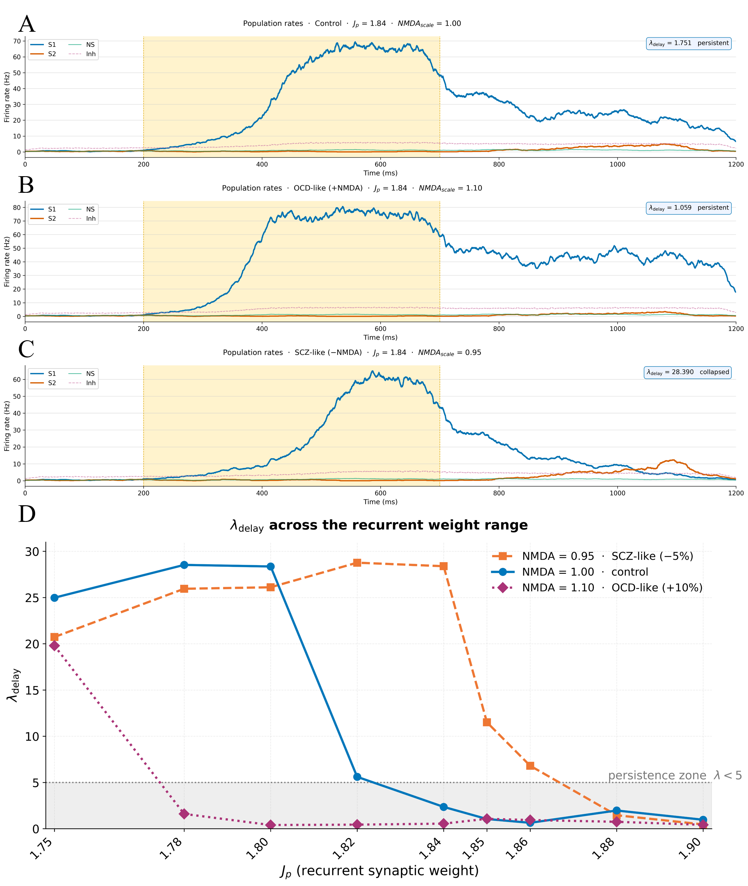
Figure 1:A--C. Representative firing-rate traces (firing rate in Hz, y-axis) of the memory-selective population (S1), competing selective population (S2), non-selective (NS) and inhibitory neurons during simulations under three NMDA scaling regimes.
(A) Control NMDA (): Healthy regime.
(B) OCD-like (): Receptor hyperfunction regime.
(C) SCZ-like (): Receptor hypofunction regime.
D. as a function of recurrent synaptic weight across the
three NMDA conditions. The shaded region marks the persistence
zone (). Conditions are distinguished by line style as well as color
to ensure accessibility.
Panels A–C of Figure 1 illustrate single-trial population rate traces for three representative conditions at . In Figure 1A (control, ), the S1 pool ramps up during stimulation and sustains elevated activity at ~20 Hz throughout the delay, indicating a successful WM maintenance. The competing pools (S2, NS) remain near baseline. In Figure 1B (OCD-like, ), the memory attractor is even more robust: activity is sustained at higher rates ~45 Hz, reflecting an over-deepened basin of attraction. In contrast, Figure 1C (SCZ-like, ) shows activity that collapses rapidly after cue offset, returning to baseline before the end of the delay, the attractor basin is too shallow to resist noise-driven escape.
Figure 1D reveals the full picture across the sweep. For the control condition, the network enters the persistence zone () at intermediate (~1.84-1.86), reflecting a tuned operating point. For the SCZ-like condition, remains elevated across nearly the entire range, until higher values of (> 1.86). For the OCD-like condition, the system drops into the persistence zone at and plummets rapidly: the network locks into persistent activity easily and at weights where the control network would still be in the non-persistent regime.
The influence of the distractor stimulus on the S2 population is modest in the current parameter regime; stronger distractor amplitudes should be explored in future work to more directly probe attractor competition and cross-pool interference.
These results are consistent with the hypothesis that NMDA conductance acts as a bidirectional gain control on attractor depth, offering a possible mechanistic link between two ostensibly opposite psychiatric presentations within a single mechanistic axis:
In schizophrenia-like regime NMDA hypofunction flattens attractor basins, making WM representations fragile, easily disrupted by noise, and prone to collapse during the delay. This is consistent with the well-documented WM deficits and hypofrontality observed in schizophrenia Goldman-Rakic, 1994Rolls, 2007, as well as the reduced signal-to-noise ratio in prefrontal circuits Winterer et al., 2004. Cognitive and negative symptoms, failure to maintain task-relevant representations and flattening of goal-directed behavior, could arise from this attractor instability.
In OCD-like regime, NMDA hyperfunction may over-deepen attractor basins, causing representations to persist beyond their usefulness and resist updating and disengagement. The network becomes locked into its current attractor state, providing a hypothetical computational analog to the perseverative thoughts and behavioral rigidity that define OCD Milad & Rauch, 2012.
Together, these simulations suggest that the healthy prefrontal WM circuit operates in a narrow, tuned window of NMDA conductance, close enough to the instability boundary to remain flexible and updatable, but deep enough to maintain representations against noise. The metric provides a continuous, quantitative readout of this balance, offering a potential link to delay-period fMRI and electrophysiology signatures.
We note that the interpretation of as a proxy for attractor basin depth is a hypothesis. Alternative mechanisms, such as a change in the effective membrane time constant of the selective population, or a shift in the competitive balance between S1 and S2, could produce similar changes in the decay timescale without necessarily reshaping the attractor landscape. Disambiguating these alternatives would require explicit computation of the flow field, as in Loh et al., 2007, and is left for future work.
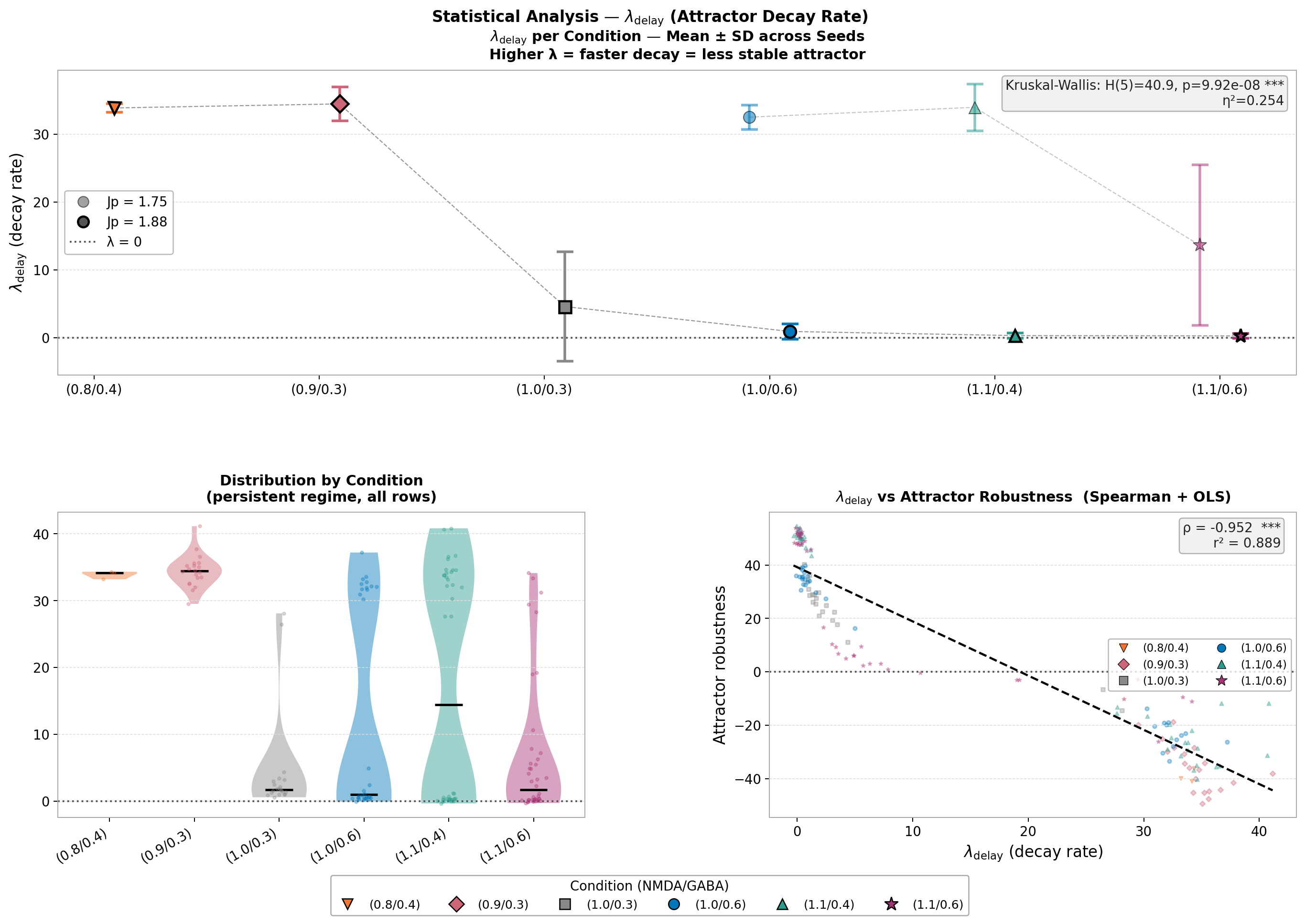
Figure 2:A. Mean ± SD of per condition across seeds for (faded) and (solid). Conditions differ significantly across the parameter space (Kruskal-Wallis: , , ).
B. Violin plots showing the full distribution of in the persistent regime across all seeds and values per condition. Black bars indicate the median.
C. Scatter plot of versus attractor robustness showing a strong Spearman correlation (, ).
To characterise attractor stability statistically across seeds and conditions, we tested different combinations of NMDA and GABA conductances at two recurrent weight values ( and ), running multiple trials with different random seeds and computing for each trial. The persistent regime () showed a median of 3.3 compared to 29.2 in the transient regime, a difference validated by a Mann-Whitney test (, ). A Kruskal-Wallis test confirmed significant differences across conditions (, , ), establishing that is sensitive to changes in NMDA/GABA conductance across the parameter space. showed a strong Spearman correlation with attractor robustness (, ), supporting its validity as a continuous measure of working memory attractor stability, capturing the same information as binary persistence classification but with greater resolution.
This work demonstrates that a single synaptic parameter, NMDA receptor conductance, is sufficient to drive a prefrontal attractor network between three qualitatively distinct regimes: healthy WM maintenance, schizophrenia-like memory collapse, and OCD-like pathological persistence. The metric provides a graded, continuous measure of this axis, moving beyond binary persistent/non-persistent classifications and offering a quantitative bridge between in silico dynamics and delay-period neural signatures measurable with fMRI or multi-unit electrophysiology. Future work should examine how dopaminergic D1 receptor modulation interacts with NMDA-mediated stability, and whether combined NMDA/GABA perturbations can reproduce the positive symptoms of schizophrenia, including spontaneous wandering between attractor states, as predicted by prior theoretical work Deco & Rolls, 2007Loh et al., 2007.
Acknowledgments
This work was supported by the Impact Scholars Program 2025. The first author is supported by the CNRST PASS doctoral scholarship program. The authors thank Archishman Biswas for his technical support with the simulation code, helpful discussions, and guidance that greatly contributed to the development of this work. The authors used AI assistance (Claude, Anthropic) to assist with scientific writing and code review during the preparation of this manuscript. All scientific content, results, and conclusions were verified and approved by the authors.
Data Availability
Published via Impact Scholars; original development repository.
- Brunel, N., & Wang, X.-J. (2001). Effects of Neuromodulation in a Cortical Network Model of Object Working Memory Dominated by Recurrent Inhibition. Journal of Computational Neuroscience, 11(1), 63–85. 10.1023/a:1011204814320
- Compte, A., Brunel, N., Goldman-Rakic, P. S., & Wang, X. J. (2000). Synaptic mechanisms and network dynamics underlying spatial working memory in a cortical network model. Cerebral Cortex, 10(9), 910–923. 10.1093/cercor/10.9.910
- Wang, X.-J. (1999). Synaptic Basis of Cortical Persistent Activity: The Importance of NMDA Receptors to Working Memory. Journal of Neuroscience, 19(21), 9587–9603. 10.1523/JNEUROSCI.19-21-09587.1999
- Wang, X.-J. (2001). Synaptic Reverberation Underlying Mnemonic Persistent Activity. Trends in Neurosciences, 24(8), 455–463. 10.1016/s0166-2236(00)01868-3
- Coyle, J. T., Tsai, G., & Goff, D. (2003). Converging Evidence of NMDA Receptor Hypofunction in the Pathophysiology of Schizophrenia. Annals of the New York Academy of Sciences, 1003, 318–327. 10.1196/annals.1300.020
- Milad, M. R., & Rauch, S. L. (2012). Obsessive-Compulsive Disorder: Beyond Segregated Cortico-Striatal Pathways. Trends in Cognitive Sciences, 16(1), 43–51. 10.1016/j.tics.2011.11.003
- Loh, M., Rolls, E. T., & Deco, G. (2007). A Dynamical Systems Hypothesis of Schizophrenia. PLOS Computational Biology, 3(11), e228. 10.1371/journal.pcbi.0030228
- Stimberg, M., Brette, R., & Goodman, D. F. M. (2019). Brian 2, an Intuitive and Efficient Neural Simulator. eLife, 8, e47314. 10.7554/eLife.47314
- Rolls, E. T., Loh, M., Deco, G., & Winterer, G. (2008). Computational models of schizophrenia and dopamine modulation in the prefrontal cortex. Nature Reviews Neuroscience, 9(9), 696–709. 10.1038/nrn2462
- Rolls, E. T., Loh, M., & Deco, G. (2008). An attractor hypothesis of obsessive–compulsive disorder. European Journal of Neuroscience, 28(4), 782–793. 10.1111/j.1460-9568.2008.06379.x
- Catts, V. S., Lai, Y. L., Weickert, C. S., Weickert, T. W., & Catts, S. V. (2016). A quantitative review of the postmortem evidence for decreased cortical N-methyl-D-aspartate receptor expression levels in schizophrenia: How can we link molecular abnormalities to mismatch negativity deficits? Biological Psychology, 116, 57–67. 10.1016/j.biopsycho.2015.10.013
- Pittenger, C., Bloch, M. H., & Williams, K. (2011). Glutamate abnormalities in obsessive compulsive disorder: Neurobiology, pathophysiology, and treatment. Pharmacology & Therapeutics, 132(3), 314–332. 10.1016/j.pharmthera.2011.09.006
- Goldman-Rakic, P. S. (1994). Working Memory Dysfunction in Schizophrenia. Journal of Neuropsychiatry and Clinical Neurosciences, 6(4), 348–357. 10.1176/jnp.6.4.348
- Rolls, E. T. (2007). Memory, Attention, and Decision-Making. Oxford University Press. 10.1093/acprof:oso/9780199232703.001.0001
- Winterer, G., Coppola, R., Goldberg, T. E., Egan, M. F., Jones, D. W., Sanchez, C. E., & Weinberger, D. R. (2004). Prefrontal Broadband Noise, Working Memory, and Genetic Risk for Schizophrenia. American Journal of Psychiatry, 161(3), 490–500. 10.1176/appi.ajp.161.3.490